How to Prepare Samples for 2D Gel Electrophoresis: A Comprehensive Step-by-Step Protocol
In the field of proteomics, 2D gel electrophoresis is a powerful and widely used technique for analyzing complex protein mixtures. This method allows for the separation of thousands of proteins based on their isoelectric point (pI) and molecular weight, providing valuable information about protein expression, post-translational modifications, and protein-protein interactions. To ensure accurate and reproducible results, the key to successful 2D gel electrophoresis lies in the proper preparation of protein samples.
In this comprehensive guide, we will walk you through a step-by-step protocol for preparing samples for 2D gel electrophoresis. This protocol is based on the practices developed by Kendrick Labs, a pioneer in the field of proteomics, and is aimed at providing a human-based, high-quality, and unique approach to achieve optimal results.
Step 1: Sample Collection and Lysis
Before starting the sample preparation, it is crucial to plan and organize the experimental design carefully. Ensure that you have sufficient biological replicates to obtain statistically meaningful results. Begin by collecting your biological samples and storing them appropriately to preserve protein integrity.
Once you have the samples, the first step is to lyse the cells or tissues to release the proteins. Homogenize the cells or tissues in a suitable lysis buffer containing detergents, protease inhibitors, and other additives to protect the proteins from degradation and maintain their native structures. The choice of lysis buffer may vary depending on the sample type and downstream applications.
Step 2: Protein Extraction
After lysis, you need to extract the proteins from the lysed cells or tissues. Centrifuge the lysate at a suitable speed and duration to pellet cellular debris and obtain the protein-containing supernatant. Transfer the supernatant to a clean tube, taking care not to disturb the pellet.
Step 3: Protein Quantification
Next, quantify the protein concentration in the extracted samples. This step is essential for normalizing the protein load in the subsequent steps of the 2D gel electrophoresis. You can use various methods for protein quantification, such as the Bradford assay, bicinchoninic acid (BCA) assay, or spectrophotometry.
Step 4: Protein Sample Preparation for 2D Gel Electrophoresis
Now that you have quantified the protein concentration, it’s time to prepare the samples for 2D gel electrophoresis. There are two main approaches to 2D gel electrophoresis: immobilized pH gradient (IPG) strips and precast gels. Let’s explore each method in detail:
Immobilized pH Gradient (IPG) Strip Method:
Rehydration of IPG Strips: Prepare the IPG strips by rehydrating them in the rehydration buffer containing the protein sample. Incubate the IPG strips for a sufficient time (e.g., 12-16 hours) to ensure optimal protein loading.
Isoelectric Focusing (IEF): Place the rehydrated IPG strips in the IEF apparatus and apply an electric field to separate the proteins based on their pI. This process may take several hours to complete. Optimize the IEF parameters, such as voltage, current, and focusing time, based on your sample characteristics.
Equilibration: After IEF, equilibrate the IPG strips in an equilibration buffer to reduce protein precipitation and prepare them for the second dimension separation.
Second Dimension Electrophoresis: Place the equilibrated IPG strips on top of the precast polyacrylamide gels and seal them with agarose. Perform SDS-PAGE (sodium dodecyl sulfate-polyacrylamide gel electrophoresis) to separate the proteins based on their molecular weight.
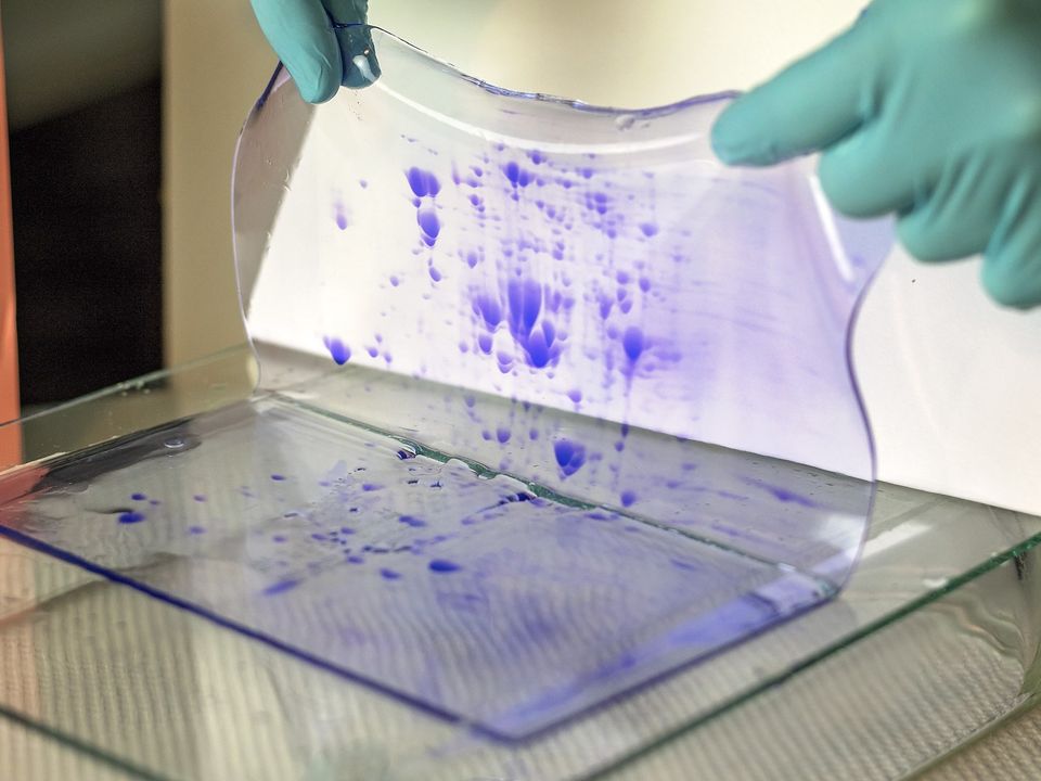
Visualization: Stain the gels with a protein stain, such as Coomassie Brilliant Blue or silver stain, to visualize the protein spots. Alternatively, for quantitative proteomics, consider using fluorescent or DIGE (difference gel electrophoresis) staining methods.
Precast Gel Method:
Sample Loading: Mix the protein sample with an appropriate sample buffer containing reducing agents and denaturants. Heat the mixture at a suitable temperature (e.g., 95°C) for a few minutes to denature the proteins.
Second Dimension Electrophoresis: Load the denatured protein samples onto the precast polyacrylamide gels and perform SDS-PAGE to separate the proteins based on their molecular weight. Visualization: Stain the gels using a protein stain, as mentioned earlier.
Step 5: Gel Imaging and Analysis
After the electrophoresis, you need to capture an image of the gels for analysis. Utilize a gel imaging system equipped with a high-resolution camera to capture the protein spots. Modern gel imaging systems offer numerous options, including fluorescence and chemiluminescence detection.
Step 6: Image Analysis and Data Interpretation
Once you have the gel images, use specialized software for image analysis and spot detection. Several commercial and open-source software packages are available for this purpose. After spot detection, compare the protein spot patterns across different gels to identify differentially expressed proteins or protein isoforms.
Conclusion
Preparing samples for 2D gel electrophoresis is a critical step that significantly influences the success of proteomic experiments. Following the step-by-step protocol provided by Kendrick Labs or any other reliable source ensures the accuracy, reproducibility, and quality of the results. By carefully collecting, lysing, and extracting proteins from your samples, quantifying the protein concentration, and choosing the appropriate method for 2D gel electrophoresis, you set a solid foundation for revealing valuable insights into the complex world of proteomics.
Proteomics continues to be an exciting and evolving field, with innovative techniques, such as mass spectrometry-based approaches, complementing 2D gel electrophoresis. However, the simplicity, cost-effectiveness, and wide application range of 2D gel electrophoresis make it an indispensable tool for proteomic researchers worldwide. Mastering the art of sample preparation for 2D gel electrophoresis is a skill that will undoubtedly yield valuable discoveries and contribute to our understanding of protein biology.